A key component to successful endodontic treatment is the early recognition of unusual canal configurations — particularly in cleaning, shaping and obturation. A recent study conducted by the Department of Conservative Dentistry and Endodontics shows the main developmental issue that causes the C-shaped root formation is the failure of the Hertwig’s epithelial root sheath to fuse to the buccal or lingual root surface. A fin or web that connects individual root canals together is the most characteristic diagnostic feature of these canals.
A traditional periapical radiograph typically is too distorted to properly identify a C-shaped canal because of the limitations of a two-dimensional image. While these canals commonly look normal at the pulp, the apical anatomy underneath it is deceivingly complex. These anomalous C-shaped canals are frequently a challenge for the dentist to properly diagnose and treat effectively.
Evidence-based facts:
- C-shaped canals were documented in 1979 by Cooke and Cox. The canals were named after the morphological cross section of the root and canal system that describes the configuration.
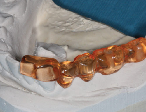
- The pulp chamber of the C-shaped canal is a single ribbon-shaped orifice with a 180° arc or more, rather than the typical discrete orifices.
- Distinct ethnic variances are highly frequent and appear most often in Asian patients with a prevalence rate of approximately 31.5% (versus 2.7–9% for Caucasians).
- C-shaped canals are found most frequently in the mandibular second molar. The prevalence rate is 2.7– 44.5%.
- The three categories of C-shaped canals are as follows: Type I is a continuous C-shaped canal; Type II is a semicolon-shaped canal; and Type III involves two or more separate canals.
- C-shaped canals characteristically are not geometrically centred in the cross section of the root, and the wall of the mesial canals is often the thinnest.
- C-shaped canals have irregular areas that can potentially contain soft-tissue remnants and/or infectious debris, which can escape during cleaning or filling, possibly causing more pain for the patient.
- The morphology of C-shaped canals varies at different levels along the length of the root. The external root surface and the internal root canal wall have far less dentinal thickness than in other teeth, so a clinical concern is that stripping of the walls may occur during shaping or post placement.