Clinical dentistry has been focusing research in the area of noninvasive and accurate diagnostic methods for the evaluation of dental pathosis. Without using ionizing radiation, the MRI has become an indispensable tool for non-invasively diagnosing and monitoring disease in soft tissues. Specific interest in this area has focused on a recently developed magnetic resonance imaging (MRI) technique called Sweep Imaging with Fourier Transform, or SWIFT. At the centre of developing studies, research of the SWIFT technology has shown great promise for advancements, especially in the field of endodontics.
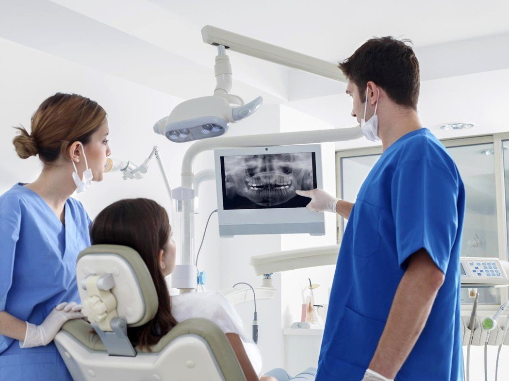
In a recent study documented in the Journal of Endodontics (June 2011) an in vivo imaging comparison was performed using SWIFT imaging system, traditional radiographs and three-dimensional cone-beam computed tomography (CBCT) scanning. The results of the research concluded that SWIFT also identified the presence and extent of dental caries and fine structures of the teeth, including cracks and accessory canals, which are not visible with existing clinical radiography techniques.
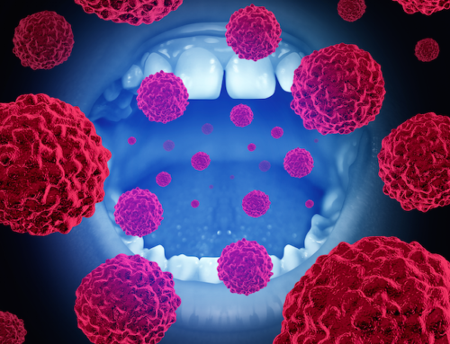
What does this mean in dentistry today? Diagnostic imaging in dentistry depends mostly on x-ray– based techniques that carry some risks and limitations such as exposure to ionizing radiation and its associated increased risk of cancer. Along with the risks, is also the inability to visualize the pulpal tissue in a consistent and detailed manner.
Although the application of x-ray– based three-dimensional diagnostic imaging is likely to increase in endodontics, as cone-beam computed tomography (CBCT) imaging systems become more available, significant limitations include both radiation as well as the inability to simultaneously image calcified and non-calcified dental tissues. This particular drawback is seen as quite a significant limitation, particularly as regenerative endodontic procedures become more common in clinical practice.
In conclusion, the SWIFT MRI offers simultaneous three-dimensional hard- and soft-tissue imaging of teeth without the use of ionizing radiation. Furthermore, it has the potential to image minute dental structures within clinically relevant scanning times. This technology has implications for endodontists due to the fact that it offers a potential method to longitudinally evaluate teeth where pulp and root structures have been regenerated as well as the ability to identify previously “invisible” structures.