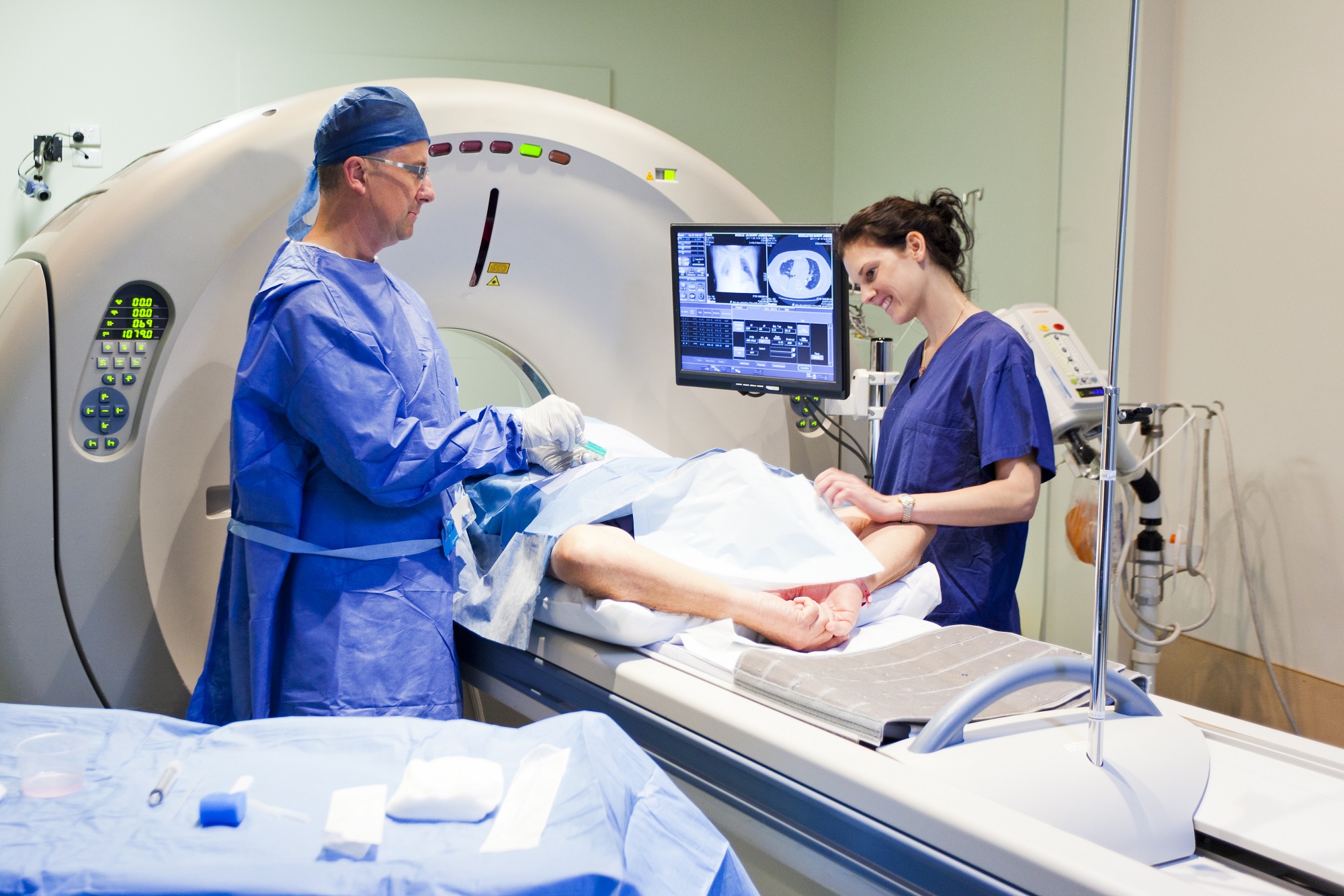
A growing body of research has demonstrated the potential for magnetic resonance imaging (MRI) in clinical dentistry, including endodontics, prosthodontics, and orthodontics. Yet there has been little analysis of how dental materials in a patient’s mouth may affect the end result, according to a new study in Dentomaxillofacial Radiology.
Contradictory results have been reported regarding the severity of image artifacts caused by different dental materials. “Magnetic susceptibility information is not readily available for many materials used in dentistry, especially those containing several components,” wrote the study authors, from the University of California, San Francisco.
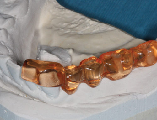
Using a 1.5-tesla MRI system and spin-echo and gradient-echo pulse sequences, the researchers investigated the potential influence of metal, ceramic, polymer, and composite dental materials on MRI. They then applied the geometric method to determine the magnetic susceptibility of the materials, and classified the materials based upon their degree of compatibility with MRI.
“The tested materials showed a range of distortion degrees,” the study authors wrote. This could be because manufacturers of these products often use iron oxide pigments, and “the smallest contamination by ferromagnetic substances can drastically alter the susceptibility of a magnetically compatible material,” researchers noted.
Julian Boldt, PhD, of the University of Würzburg and co-author on the Dentomaxillofacial Radiology study, says he and his colleagues are close to completing a prototype of a dedicated dental MRI scanner with the potential for therapeutic applications in dentistry, such as digital impressions. His team recommends regulatory standards requiring the magnetic susceptibility value limit for dental materials be within quantifiable magnetic compatibility limits.