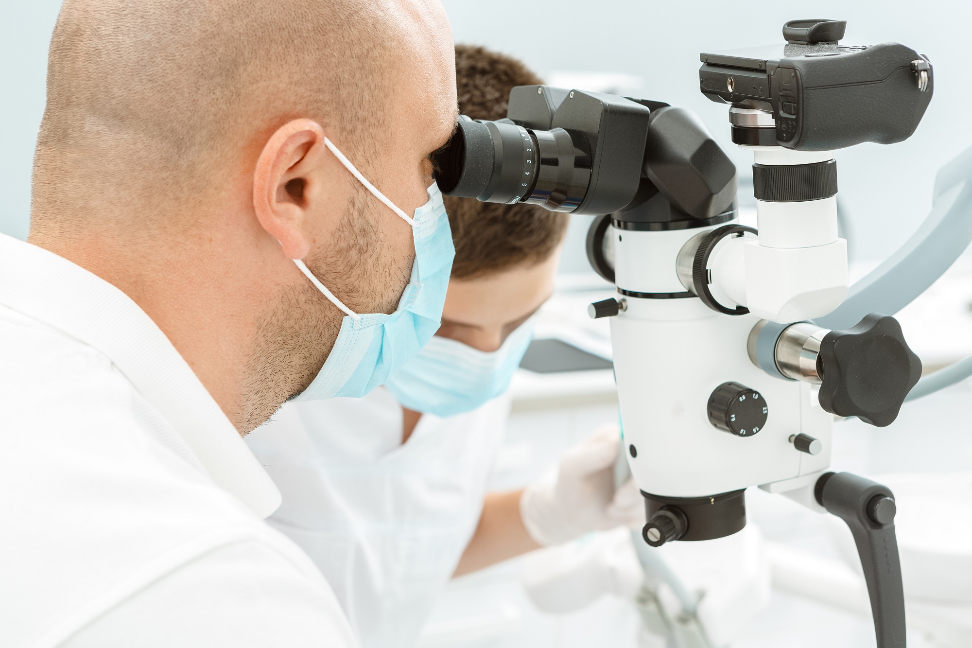
Surgical and non-surgical endodontics has been completely transformed by the use of illuminated dental microscopes in clinical procedures. For many years, surgical burs and amalgam for root-end fillings were the standard of care. Now with the incorporation of the operating microscope, and also the endoscope, along with the use of ultrasonic tips and biocompatible filling materials, there is an emerging new standard of care in modern endodontic microsurgery.
The steps of endodontic microsurgery are now carried out with varying degrees of magnification, including flap preparation, osteotomy identification of root apices, root-end resection, inflammatory tissue removal, observation of the resected root surface, root-end preparation, root-end filling, and suturing. The microscope is also helpful for cervical or external resorption or perforation repairs.
An operating microscope also aids in caries removal, access preparation, removal of pulp chamber calcifications and the identification of root canal orifices. An endodontist is better able to detect subtle changes in dentin colour and texture. Developmental lines on the pulp floor guide the practitioner towards root canal orifices, allowing safer dentin removal.
Microscope magnification helps in the instrumentation of obstructed and calcified canals, canal bifurcations and canal obstructions such as denticles, calcifications and obturation. Additional primary endodontic procedures that benefit from microscope use include vital pulp therapy and regenerative endodontics by allowing careful and gentle manipulation of the pulpal tissues. The microscope is helpful in identifying and removing leftover filling materials, such as sealer remnants, pastes or gutta percha, silver points and carrier-based materials. Microscopes also aid in nonsurgical perforation repair, allowing the practitioner to clean the perforation site and place the repair material more precisely.
The dental operating microscope is now considered an integral part of endodontic practice. For both nonsurgical and surgical endodontic treatment, use of microscopes results in improved outcomes for patients, and current endodontic therapy is demonstrably more effective because of its use.