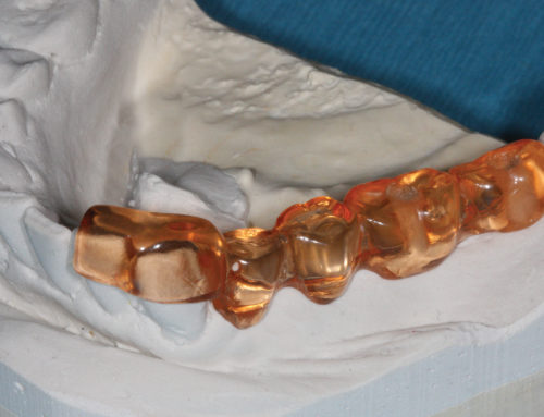

Endodontic treatment often has success rates of 90-95%. Yet, despite well-done initial root canal treatment, some cases fail and require retreatment. Endodontists now have technologically enhanced instruments, like the dental operating microscope and 3D cone-beam imaging, which allow for a more precise diagnosis of a failing root canal, thereby facilitating retreatment of the case.
The dental operating microscope and 3D cone-beam imaging improve on conventional visual evaluations of teeth and the surrounding tissues. The advanced technology vastly improves the visualization of apical pathology and offers far more nuanced clinical treatment choices, thereby preventing both under-treatment and over-treatment. An endodontist can confirm the exact nature of the issue that requires retreatment and, often, may prevent root amputation or extraction.
Dental Operating Microscope: This indispensable endodontic clinical tool provides enhanced magnification that allows unparalleled visual acuity of the tooth structure that is being restored and precise views of internal anatomy, clinical endo access and the pulp chamber. Pathology is now clinically visible, which can determine whether a fracture exists. It can also identify whether a canal is untreated and/or calcified, which is often associated with etiologies in a failing root canal. The quality of endodontic care is improved, and outcomes are far more predictable.
3D Cone-Beam Imaging: Conventional 2D radiograph imaging, like periapicals and panoramic radiographs, are limited in the clinical information provided to an endodontist found in a retreatment clinical scenario. Previously treated root canals that utilized 2D radiographs for initial treatment often reveal unseen apical pathology or anatomical anomalies not seen in the traditional radiographs when reevaluated with 3D cone-beam imaging. A 3D scan enables the determination of the exact location of the endo-related pathology.
Endodontists no longer need to make educated guesses when managing root canal retreatment cases. The dental operating microscope and 3D cone-beam imaging allow more conservative and less invasive treatment options for dentists and their patients.