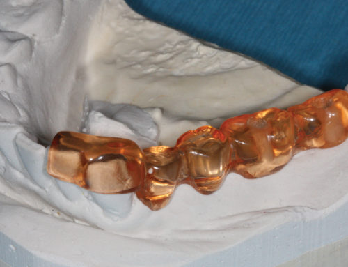
Of all dental specialties, endodontics may have realized the most benefits of technological advancements. A trending paradigm that preservation of tooth structure may be a better predictor of long-term outcomes is supported by Ellen Meyer, DMD, who in Inside Dentistry writes, “Creating more conservative access openings can reduce the risk of tooth loss related to weakened tooth structure. Significant problems are more related to the loss of tooth structure more than the endodontic procedures themselves.”
So, what factors drive the implementation of a minimally invasive approach? Three pieces of technology have allowed this paradigm shift in endodontic practice: the dental operating microscope, root-form-heattreated NiTi instruments with placement bend properties to rotary files and, importantly, the focused-field CBCT scanner. This triad of factors supports the clinical implementation of a more conservative approach to coronal enlargement and the prevention of instrument fracture.
The dental operating microscope allows more precise access, which is dramatically smaller than the traditional access to a canal. The precision can be individualized per root or even per canal. The primary objection to this smaller access is that of the limitations on the clinician’s ability to locate relevant anatomy. The impetus to use minimally invasive, restoratively driven treatment strategies is lessened with diminished visual acuity.
The advent of root-form-appropriate, variable taper files offers safe and effective instrumentation and prevents unnecessarily weakening tooth structure. Again, the primary clinical objection is that a smaller shape may limit a dentist’s ability to cleanse and shape the root canal under treatment.
The last crucial element is the use of a CBCT scanner to complement the dental operating microscope so that access can be extended if necessary but conservatively managed during treatment. CBCT imaging is now considered a requirement of a state-of-the-art endodontic practice. The utilization of CBCT imaging supports the minimization of access extension because the distance and direction are known from the imaging study.
Access to the complex anatomy of the root canal system is the most important phase of any nonsurgical root canal procedure. An endodontist is already treating a compromised tooth. Therefore, the importance of minimally invasive access openings to save existing tooth structure cannot be overstated.


