Is today’s endodontic therapy best practice to obturate a root canal to the radiographic apex? Is anything short of that radiographic apex unacceptable?
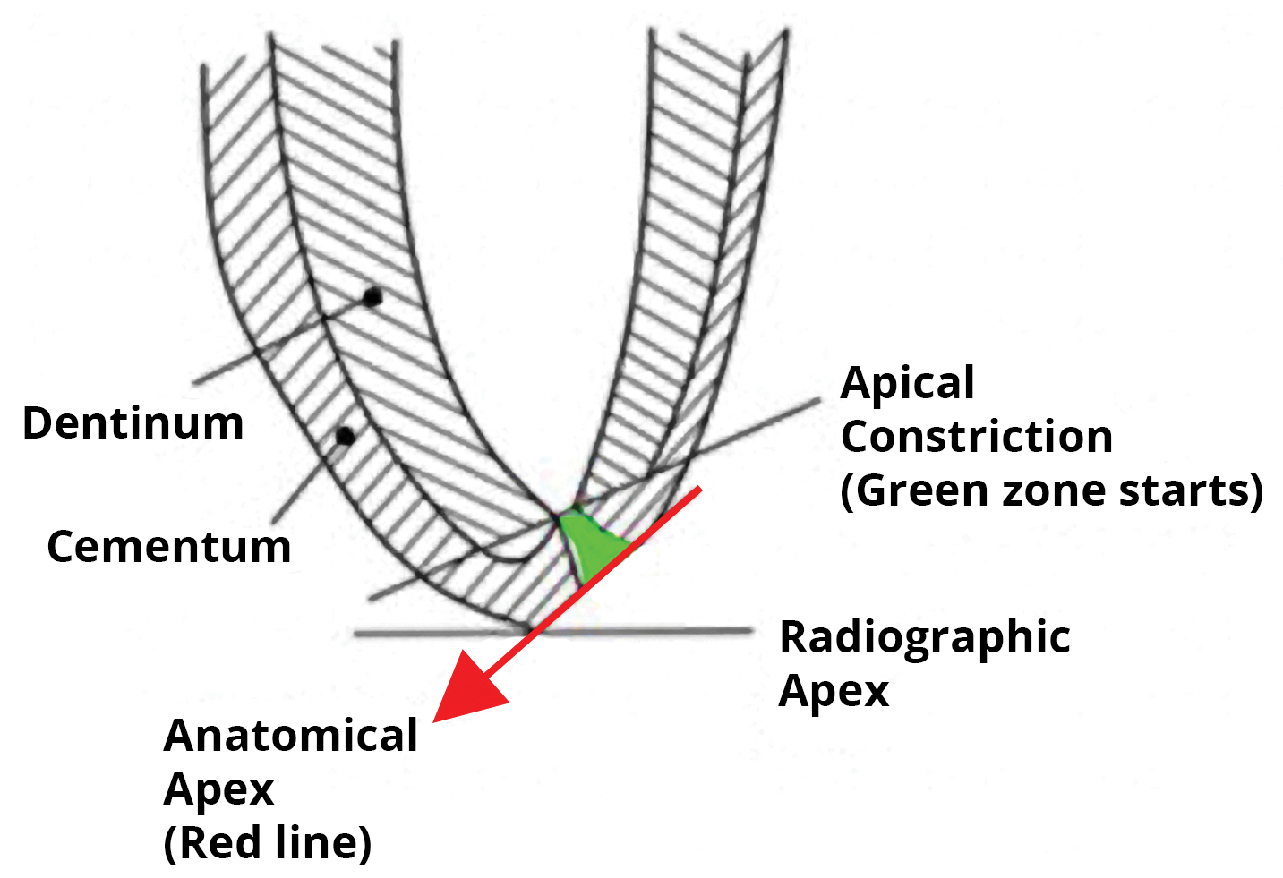
While some dental institutions still instruct that the root canal should be obturated to the radiographic apex, other clinical opinions differ on this subject. The anatomic reality and radiographic perceptions/limitations are more complex. New research indicates that a better outcome rate is achieved when treatment includes obturation short of the radiographic apex.
So, if short of the radiographic apex is desired, just how short is the goal? Published studies, dating from 2001 to the present, can help guide an informed clinical opinion. For example, research by Kojima et al. stated that the root canal should be filled to within 2 mm of the radiographic apex. Ng et al. found that other clinical considerations, including root filling extending to 2 mm within the radiographic apex, realized significant outcome improvements as related to the primary root canal treatment.
In a more recent study by Peak, Bryant and Dummer, the use of cold lateral condensation of gutta-percha to within 2 mm of the radiographic apex of the tooth was associated with the best outcomes. Chanda reported that an important factor that significantly favoured endodontic success was root filling to 2 mm within the radiographic apex and not beyond. Gomes et al. conducted a large study of 1,290 root canal teeth. The analysis showed that canals filled up to 0 mm to 2 mm short of the apex had significantly higher numbers of teeth scoring as healthy when compared with overfilled or under-filled cases.
In all of these many studies, the recurring number for a higher success rate appears to be a root canal filling ending between 0 mm to 2 mm short of the radiographic apex.
References: Kojima K, et al. (2004), Ng YL, et al. (2008), Peak JD, et al. (2009), Chanda A, (2009), Gomes AC, et al. (2015)